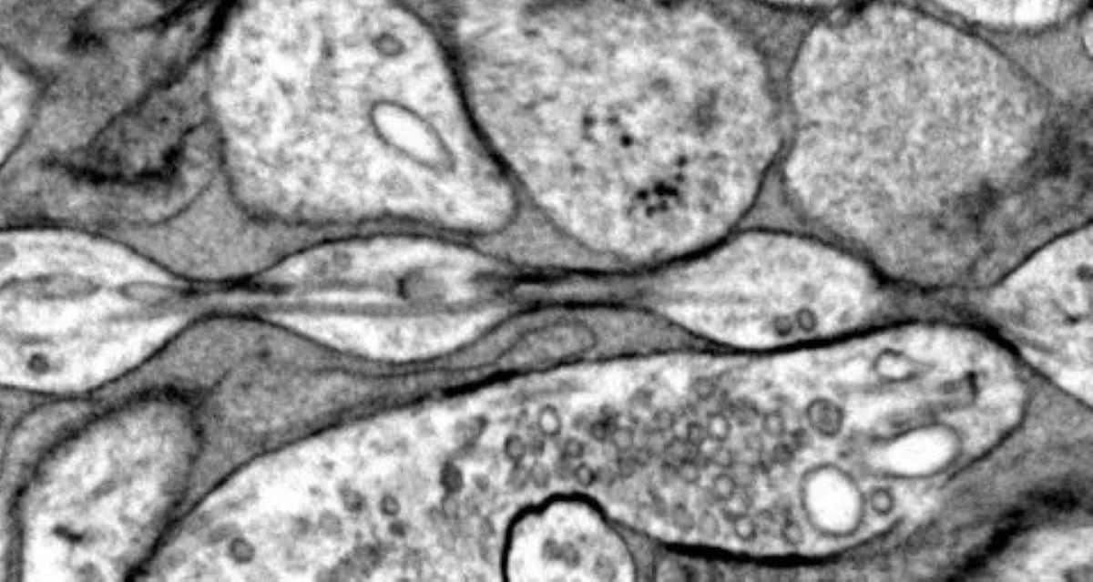
Summary: A brand new examine challenges the long-held perception that axons, mind cell extensions, are tube-like, revealing as an alternative a “pearl-on-a-string” construction.
Using superior freezing electron microscopy, researchers discovered these bead-like formations, termed “non-synaptic varicosities,” throughout mouse neurons. These constructions could affect mind signaling by modulating ion circulation and electrical sign velocity. Changes in membrane stiffness, akin to lowered levels of cholesterol, alter the pearls’ measurement and sign transmission capability.
This discovery reshapes our understanding of neuron construction and opens doorways for exploring its implications in neurodegenerative ailments. Future work will look at these formations in human mind tissues.
Key Facts:
- Axons exhibit “pearl-on-a-string” shapes, not fixed cylindrical tubes.
- Removing ldl cholesterol in axons reduces pearling and slows electrical indicators.
- High-frequency electrical stimulation briefly swells these pearl constructions.
Source: Johns Hopkins Medicine
Biology textbooks may have a revision, say Johns Hopkins Medicine scientists, who current new proof that an armlike construction of mammalian mind cells could also be a distinct form than scientists have assumed for greater than a century.
Their examine on mouse mind cells reveals that the cells’ axons — the armlike constructions that attain out and alternate data with different mind cells — should not the cylindrical tubes usually pictured in books and on web sites however extra like pearls on a string.

A report on the findings is printed on-line Dec. 2 in Nature Neuroscience.
“Understanding the construction of axons is essential for understanding mind cell signaling,” says Shigeki Watanabe, Ph.D., affiliate professor of cell biology and neuroscience on the Johns Hopkins University School of Medicine. “Axons are the cables that join our mind tissue, enabling studying, reminiscence and different capabilities.”
Scientists have recognized that pearl-like constructions in axons, known as axon beading, can develop in dying mind cells and in individuals with Parkinson’s and different neurodegenerative ailments as a result of lack of membrane and skeletal integrity in neurons.
Under regular situations, axons are considered formed like tubes with a largely fixed diameter and occasional bubble-like constructions (synaptic varicosities that maintain globs of neurotransmitters, which allow signaling to different mind cells).
Watanabe had initially seen repeated axon pearling within the nervous system of worms and grew extra curious concerning the constructions after a dialogue with Swiss scientist Graham Knott, Ph.D. A analysis staff from Harvard University had printed a examine in 2012 that recognized repeated “skeletal” elements in axons, so the pair of researchers mentioned experiments to eliminate the axon skeleton to see if the pearl constructions disappear, says Watanabe.
Johns Hopkins graduate scholar and examine first creator Jacqueline Griswold examined the thought however discovered no impact on axon pearling.
Then, Watanabe and Griswold labored with theoretical biophysics colleague Padmini Rangamani, Ph.D., professor of pharmacology at University of California San Diego School of Medicine, to look extra intently at axons’ bodily properties.
To be capable to see axons on mind cells (neurons), that are 100 instances smaller than the width of a human hair, the scientists used excessive strain freezing electron microscopy.
Like normal electron microscopy, which shoots beams of electrons at a cell to stipulate its construction, Watanabe and his staff froze mouse neurons to protect the constructions’ form.
“To see nanoscale constructions with normal electron microscopy, we repair and dehydrate the tissues, however freezing them retains their form — just like freezing a grape relatively than dehydrating it right into a raisin,” says Watanabe.
The researchers studied three forms of mouse neurons: ones grown within the lab, these taken from grownup mice and people taken from mouse embryos. The neurons had been nonmyelinated (they had been with out the myelin insulating cowl that surrounds the axon).
The researchers discovered the bubbly, pear form of axons amongst all the tens of 1000’s of photos taken of the tissue samples.
The scientists named the pearl-like constructions during which the axon swells “non-synaptic varicosities.”
“These findings problem a century of understanding about axon construction,” says Watanabe.
The scientists additionally used mathematical modeling to see if the axon membrane influenced the form or presence of the pearl on a string construction. They discovered that easy mechanical fashions might be used to elucidate these constructions very successfully.
Furthermore, experiments with the mathematical mannequin and mouse mind samples confirmed that growing the focus of sugars within the resolution across the axon or lowering pressure within the axonal membranes lowered the pearl constructions’ measurement.
In one other experiment, the scientists eliminated ldl cholesterol from the neuron’s membrane to make it much less stiff and extra fluid-like. Under this situation, they discovered much less pearling in each mathematical fashions and mouse neurons, together with lowered capability of the axon to transmit electrical indicators.
“A wider area within the axons permits ions [chemical particles] to go by way of extra rapidly and keep away from visitors jams,” says Watanabe.
The scientists additionally utilized excessive frequency electrical stimulation to the mouse neurons, which made pearled constructions alongside axons swell, on common, 8% longer and 17% wider for a minimum of 30 min after stimulation and elevated the velocity {of electrical} indicators.
However, when ldl cholesterol was faraway from the membrane, the axon’s pearls misplaced their swollen state and had no change within the velocity {of electrical} indicators.
The analysis staff plans to look at axonal “arms” in human mind tissue taken with permission from individuals having mind surgical procedure and people who have died from neurodegenerative ailments.
This work fashioned the premise of a not too long ago awarded Multiple Principal Investigator grant to Watanabe and Rangamani from the National Institute of Mental Health.
Funding: Funds for the analysis had been offered by the Johns Hopkins University School of Medicine, the Marine Biological Laboratory Whitman Fellowship, the Chan Zuckerberg Initiative Collaborative Pair Grant and Supplement Award, the Brain Research Foundation Scientific Innovations Award, a Helis Foundation award, the National Institutes of Health (NS111133-01, NS105810-01A11, DA055668-01, 1RF1DA055668-01), the Air Force Office of Scientific Research (FA9550-18-1-0051), the Alfred P. Sloan Research Fellowship, a McKnight Foundation scholarship, a Klingenstein-Simons Fellowship Award in Neuroscience, a Vallee Foundation scholarship, the National Science Foundation and the Kavli Institutes at Johns Hopkins and UC San Diego.
Other researchers who performed the examine are Chintan Patel, Renee Pepper, Sumana Raychaudhuri, Quan Gan, Sarah Syed and Brady Maher from Johns Hopkins, Mayte Bonilla-Quintana, Christopher Lee, Cuncheng Zhu and Miriam Bell from the UC San Diego, Siyi Ma from the Marine Biology Laboratory, Mitsuo Suga and Yuuki Yamaguchi from JEOL in Tokyo, and Ronan Chéreau and U. Valentin Nägerl from the Université de Bordeaux in France.
About this neuroscience analysis information
Author: Vanessa Wasta
Source: Johns Hopkins Medicine
Contact: Vanessa Wasta – Johns Hopkins Medicine
Image: The picture is credited to Quan Gan, Mitsuo Suga, Shigeki Watanabe
Original Research: Open entry.
“Membrane mechanics dictate axonal pearls-on-a-string morphology and performance” by Shigeki Watanabe et al. Nature Neuroscience
Abstract
Membrane mechanics dictate axonal pearls-on-a-string morphology and performance
Axons are ultrathin membrane cables which can be specialised for the conduction of motion potentials. Although their diameter is variable alongside their size, how their morphology is set is unclear.
Here, we show that unmyelinated axons of the mouse central nervous system have nonsynaptic, nanoscopic varicosities ~200 nm in diameter repeatedly alongside their size interspersed with a skinny cable ~60 nm in diameter like pearls-on-a-string. In silico modeling means that this axon nanopearling may be defined by membrane mechanical properties.
Treatments disrupting membrane properties, akin to hyper- or hypotonic options, ldl cholesterol removing and nonmuscle myosin II inhibition, alter axon nanopearling, confirming the function of membrane mechanics in figuring out axon morphology.
Furthermore, neuronal exercise modulates plasma membrane ldl cholesterol focus, resulting in modifications in axon nanopearls and inflicting slowing of motion potential conduction velocity.
These knowledge reveal that biophysical forces dictate axon morphology and performance, and modulation of membrane mechanics seemingly underlies unmyelinated axonal plasticity.